
Review | Open Access | Volume 9 (4): Article 198 | Published: 22 Nov 2026

Views: 39
Menu, Tables and Figures
Table 1: Epidemiology of Leishmaniasis in Sub-Saharan African Countries (2010–2025)
| S/No | Country | Prevalence | Type of Study | Year of Data Collection | Population Type | Location | Age | Type of Leishmaniasis | Species of Leishmania | Reference |
|---|---|---|---|---|---|---|---|---|---|---|
| 1 | Burkina Faso | 44.0% parasitological positivity among suspected cases; 96 confirmed cases | Retrospective hospital case series | 2012–2016 | Patients with suspected CL | Ouagadougou | ~30.8 years average among those with age reported | CL | Leishmania major | [12] |
| 2 | Ghana | 31.9% among persons with skin ulcers detected by PCR; 41.8% exposure by leishmanin skin test | Cross-sectional community surveys | 2018 | Community residents and persons with skin ulcers | Oti Region (Ashiabre, Keri, Sibi Hilltop) | Not reported | CL | Leishmania spp. (PCR positive) | [7] |
| 3 | Mali | LST positivity 85.1% (Diema), 24.6% (Kolokani), 2.7% (Kolondieba) with PCR-confirmed lesions in some cases | Population survey using leishmanin skin test and PCR confirmation | 2012–2016 (community survey) | General community residents in multiple districts | Districts: Diema, Kolokani, Kolondieba | Median age: 23 years | CL | Leishmania major | [9] |
| 4 | Mauritania | 0.23% | Prospective multicenter descriptive epidemiological and clinico-biological study | 2016–2017 | Patients presenting with suspected cutaneous leishmaniasis lesions at two public hospitals in Nouakchott | Nouakchott | 23 years | CL | Leishmania major in 61.9% of cases; Leishmania infantum detected in 23.8% of cases | [24] |
| 5 | Niger | 56.0% (33 confirmed CL among 59 suspected) | Clinic case series | 2023 | Patients with suspected CL lesions | Zinder area | Not reported | CL | Leishmania major | [10] |
| 6 | Nigeria | 85.7% (6/7 suspected with molecular confirmation of CL lesions) | Molecular case series | 2024 | Patients with suspected CL lesions | Dutsin-Ma LGA (Katsina State) | Not reported | CL | Leishmania major | [11] |
| 7 | Senegal | 38 cases reported | Retrospective hospital case series | 2010–2014 | Patients with CL presenting to dermatology department | Aristide Le Dantec University Hospital Dermatology, Dakar | ~25 years | CL | Not specified | [25] |
| 8 | Ethiopia | DAT – 12.5%; Seroprevalence – 32.5% | Cross-sectional study | October 2013 | Labour migrant workers | Kafta-Humera lowlands, Northwest Ethiopia | Range 14–61 years (79.9% aged 15–34) | Visceral | Leishmania donovani | [3] |
| 9 | Ethiopia | 21.6% | Cross-sectional study | January 2014–June 2014 | Clinically suspected VL patients, predominantly males (87%), including age groups under 5 years and 15–29 years, with farmers (69.5%) and rural residents (60.9%) | Metema Hospital, North Gondar Administrative Zone, Amhara region, Northwest Ethiopia | 28.1 years (SD 11.8, range 2–78 years) | Visceral | Leishmania donovani | [27] |
| 10 | Ethiopia | 9.6% | Community-based cross-sectional study | October–December 2016 | Migrant laborers aged 15–65, predominantly 15–29 years (74.2%), from lowland areas (61.2%), engaged in sesame and sorghum harvesting | Agricultural camps, West Armachiho district, Northwest Ethiopia | 15–65 years | Visceral | Leishmania donovani | [30] |
| 11 | Ethiopia | 12.8% in males and 4.2% in females | Prospective cohort | 2015–2016 | HIV-infected individuals | Metema District Hospital, North-West Ethiopia | Median 38 years (IQR 30–45) | Visceral | Leishmania donovani | [29] |
| 12 | Ethiopia | 43.3% | Retrospective cohort study | September 2013–September 2018 | VL patients, including children (51.6%), with concurrent conditions (e.g., severe acute malnutrition 27.1%, tuberculosis 6.4%, malaria 6.4%, HIV 1.1%) | Negelle General Hospital, Yabello General Hospital, Ginnir General Hospital, Oromia region, Ethiopia | Not reported | Visceral | Leishmania donovani | [28] |
| 13 | Ethiopia | 3.0% (first comers at T1), 22.1% (repeat comers at T1), 34.8% (first comers at T2), 36.4% (repeat comers at T2) | Prospective cohort | July–August 2019 (T1) to November 2019–January 2020 (T2) | Migrant workers | West Armachiho district, Northwest Ethiopia | Median 20 years | Visceral (asymptomatic) | Leishmania donovani | [31] |
| 14 | Uganda | 5.21% (95% CI: 3.15%–8.48%) in Moroto district, with county-level prevalence of 6.90% in Matheniko, 4.49% in Tepeth, and 3.61% in Moroto Municipality | Cross-sectional study | Not reported (published 2025) | General community population | Moroto district, Northeastern Uganda | Not reported | Visceral | Not reported | [6] |
| 15 | Ethiopia | Not reported (outbreak with 1050 cases) | Cross-sectional study | 2023 | Male militia members | Somali Region, Eastern Ethiopia | Not reported | Cutaneous | Leishmania tropica | [32] |
| 16 | Sudan | VL/HIV co-infection: 3.8%; VL/HBV co-infection: 1.9%; VL/HCV co-infection: 1.3%; VL/Malaria co-infection: 9.3%; VL/HIV/HBV/HCV/Malaria co-infection: 1.9% | Retrospective, hospital-based study | January 2013–June 2014 | Confirmed VL patients, predominantly males (75.7%), farmers (56.2%), rural residents (74.4%) | Gedarif Teaching Hospital, Eastern Sudan | 31.4 years | Visceral | Leishmania donovani | [33] |
| 17 | Kenya | 80.4% | Secondary data analysis and frequency-matched case-control study | 1st January 2014–11th July 2017 | Confirmed VL cases (83% males) and controls (age-matched, VL-negative), including children under 5 years | Marsabit County Referral Hospital and Laisamis Mission Hospital, Northern Kenya | Not reported | Visceral | Not specified | [34] |
| 18 | Zambia | ~7.0% | A metagenomic next-generation sequencing-based pilot study was conducted on bio-banked nasopharyngeal samples | November 2020–February 2021 | COVID-19 referral centers in Lusaka, Zambia, the University Teaching Hospital (UTH) and Levy Mwanawasa University Teaching Hospital (LMUTH) | Lusaka | ≥18 years | Molecular detection / Exposure (Nasopharyngeal mNGS) | L. braziliensis | [40] |
Note: This table summarizes population-level epidemiological surveys, hospital cohorts, and community-based molecular studies. Singular clinical case reports and isolated travel-associated infections have been excluded from this summary to maintain comparability across prevalence data. CL – cutaneous leishmaniasis; VL – visceral leishmaniasis; DAT – direct agglutination test; HBV – hepatitis B virus; HCV – hepatitis C virus.
Table 1: Epidemiology of Leishmaniasis in Sub-Saharan African Countries (2010–2025)
Table 2: Recommended treatment regimens for leishmaniasis in Sub-Saharan Africa
| Clinical Form | Primary Regional Focus | First-Line Recommended Treatment | Second-Line / Alternative Treatment |
|---|---|---|---|
| Visceral Leishmaniasis (VL) | East Africa | Sodium Stibogluconate (SSG) + Paromomycin (PM) combination | Liposomal Amphotericin B (L-AmB) or Miltefosine |
| Cutaneous Leishmaniasis (CL) – Uncomplicated | West & Southern Africa | Local therapy (Intralesional antimonials, cryotherapy, or thermotherapy) | Systemic antimonials or oral Fluconazole |
| Cutaneous Leishmaniasis (CL) – Complex/Multiple lesions | Cross-regional | Systemic antimonials (SSG or Meglumine antimoniate) | Miltefosine or Liposomal Amphotericin B (L-AmB) |
Table 2: Recommended treatment regimens for leishmaniasis in Sub-Saharan Africa
Adedayo Oluwadamilola Adesida1,2, Blessing Mercy Babalola1,2, Praise Irabor2, Tobi Emmaneul Eludipo1,2, Oyetunde Timothy Oyeyemi1,2, &
1Department of Biosciences and Biotechnology, University of Medical Sciences, Ondo, Ondo State, Nigeria, 2Tropical Medicine & Diagnostics Development Group, Seeding Graduate Laboratory, University of Medical Sciences, Ondo, Ondo State, Nigeria
&Corresponding author: Oyetunde Timothy Oyeyemi, Department of Biosciences and Biotechnology, University of Medical Sciences, Ondo City, Ondo State, Nigeria, Email: ooyeyemi@unimed.edu.ng, ORCID: https://orcid.org/0000-0002-3089-2578
Received: 05 Jun 2026, Accepted: 19 Nov 2026, Published: 22 Nov 2026
Domain: Neglected Tropical Diseases
Keywords: Leishmaniasis, epidemiology, coinfection, diagnostics, sub-Saharan Africa
©Adedayo Oluwadamilola Adesida et al. Journal of Interventional Epidemiology and Public Health (ISSN: 2664-2824). This is an Open Access article distributed under the terms of the Creative Commons Attribution International 4.0 License (https://creativecommons.org/licenses/by/4.0/), which permits unrestricted use, distribution, and reproduction in any medium, provided the original work is properly cited.
Cite this article: Adedayo Oluwadamilola Adesida et al., Leishmaniasis in sub-Saharan Africa: Evolving epidemiology, emerging transmission foci, and syndemic complexity. Journal of Interventional Epidemiology and Public Health. 2026; 9(4):198. https://doi.org/10.37432/jieph-d-26-00176
Leishmaniasis remains a neglected tropical disease with a complex and under-recognized epidemiology in Sub-Saharan Africa (SSA). This review synthesizes current evidence on the distribution, transmission dynamics, species diversity, coinfections, diagnostics, and control challenges of leishmaniasis across the region. Epidemiological data reveal marked heterogeneity, with cutaneous leishmaniasis (CL), predominantly caused by Leishmania major, widely distributed in West Africa, often exhibiting high focal prevalence. In contrast, visceral leishmaniasis (VL), driven mainly by Leishmania donovani, is concentrated in East Africa and the Horn of Africa, where prevalence is amplified by migration, occupational exposure, and ecological factors. Emerging evidence challenges previous assumptions of limited transmission in Southern Africa, suggesting underreporting linked to weak surveillance systems. Coinfections, particularly VL-HIV, as well as malaria, tuberculosis, and malnutrition, significantly worsen disease outcomes and complicate diagnosis and treatment. Diagnostic approaches have evolved from conventional microscopy to include serological and molecular techniques; however, access and performance remain uneven across endemic settings. Advances in molecular biology have enhanced understanding of parasite diversity, virulence, and drug resistance mechanisms, highlighting implications for treatment failure and control strategies. Therapeutic options remain limited, with increasing reliance on combination therapies, drug repurposing, and nanotechnology-based delivery systems. Overall, leishmaniasis in SSA is more widespread and operationally challenging than previously appreciated. Strengthening surveillance, improving diagnostic accessibility, addressing coinfections, and integrating molecular insights into control programs are critical to advancing effective and sustainable interventions in the region.
Leishmaniasis is a vector-borne neglected tropical disease caused by protozoa of the genus Leishmania and transmitted by phlebotomine sandflies. Globally, the disease presents three forms, including visceral (VL), cutaneous (CL), and mucocutaneous. The VL forms can be deadly if not treated. The disease is closely linked to poverty, poor nutrition, displacement, and a weak health system, with an estimated 700,000 to 1 million new cases occurring annually across tropical and subtropical regions [1,2]. Despite sustained global attention, the epidemiology of leishmaniasis is grossly underreported, and its true burden is challenged by inadequate surveillance, limited diagnostic options and access, and frequent misclassification with other febrile or dermatological conditions.
Within this global context, the epidemiological landscape of leishmaniasis in Sub-Saharan Africa (SSA) is among the most complex. Countries in East Africa, particularly Ethiopia, Sudan, Kenya, and Uganda, remain a major focus for VL caused predominantly by Leishmania donovani. Community surveys, hospital cohorts, and migrant worker studies show prevalence ranging from 5% in general populations to over 40% in confirmed VL cohorts, alongside substantial reservoirs of asymptomatic infection that sustain transmission[3–6]. Outbreaks linked to labour migration, militia deployment, and rural occupational exposure further complicate control.
Historically, the epidemiological relevance of leishmaniasis in West Africa is considered minor; nonetheless, the region is now recognized as harbouring active CL transmission driven largely by Leishmania major. Molecular and parasitological studies in Burkina Faso, Ghana, Mali, Mauritania, Niger, Nigeria, and Senegal reveal high positivity rates among suspected cases, often exceeding 40–80%, demonstrating established endemic foci previously undocumented in regional maps of transmission [7–12].
Southern Africa has long been regarded as largely free of leishmaniasis; however, emerging reports of human CL in Angola after decades of silence and travel-associated infections linked to South Africa and Botswana suggest that this perception reflects surveillance failure rather than true absence of disease [13–15].
A critical feature of leishmaniasis epidemiology in SSA is its interaction with other endemic diseases and comorbid conditions. VL-HIV coinfection is especially prominent in Ethiopia and Sudan, where 20–40% of VL patients in some endemic zones are HIV-positive, leading to atypical clinical presentations, high relapse rates, altered drug pharmacokinetics, and elevated mortality [2,16–18]. Coinfections with malaria and tuberculosis, alongside widespread malnutrition, further complicate diagnosis, treatment outcomes, and control efforts, embedding leishmaniasis within a broader syndemic framework [19,20].
Control efforts across SSA are hindered by diagnostic limitations, weak surveillance systems, poor species identification, limited access to effective therapeutics, vector ecology knowledge gaps, and socioeconomic drivers such as migration, conflict, and poverty. Fortunately, the recent development and application of advanced molecular and diagnostic methodologies offer promising new avenues for improving disease detection and regional surveillance [21–23].
Despite sustained global attention, critical knowledge gaps continue to obscure the true burden of leishmaniasis in Sub-Saharan Africa. Specifically, transmission dynamics in non-traditional endemic zones (such as Southern Africa) remain poorly mapped due to weak surveillance; the synergistic drivers of leishmaniasis coinfections (particularly with HIV, malaria, and tuberculosis) are not fully understood within a syndemic framework; and there is a persistent disconnect between advanced molecular discoveries—such as genomic markers of drug resistance—and their practical application in field-level control programs.
To address these gaps, this review synthesizes current knowledge on the Sub-Saharan African epidemiology of leishmaniasis by integrating evidence on species distribution, transmission ecology, and emerging geographic foci. It further examines the burden imposed by syndemic coinfections and comorbidities, evaluates existing diagnostic approaches, and bridges molecular insights with public health strategies to provide a coherent evidence base for enhanced leishmaniasis control initiatives across the region.
Search strategy and selection criteria
We approached this comprehensive literature review systematically to ensure a robust and well-rounded synthesis. We identified relevant studies by searching PubMed, Scopus, and Google Scholar. Our search relied on a combination of primary keywords and Medical Subject Headings (MeSH), specifically: (“Leishmaniasis” OR “visceral leishmaniasis” OR “cutaneous leishmaniasis”) AND (“Sub-Saharan Africa” OR “West Africa” OR “East Africa” OR “Southern Africa” OR “Central Africa”)
To ensure the review reflects the most current epidemiological landscape, the search—conducted during the January–March 2026 window—was limited to articles published between January 2010 and December 2025. We included peer-reviewed research, epidemiological surveys, hospital case series, and molecular studies published in English. Studies were included if they directly addressed human Leishmania infections, transmission vectors, coinfections, or diagnostics within Sub-Saharan Africa. Studies focused entirely on non-African epidemiological settings were excluded for regional prevalence estimations, though select non-African studies were referenced comparatively for fundamental biological, structural, or methodological principles. As this was a narrative rather than a systematic review, no formal screening counts, duplicate-removal log, or pooled quantitative analysis were undertaken.
Evidence synthesis
The identified literature was synthesised narratively and organised thematically into regional epidemiology and species distribution; transmission determinants and risk patterns; coinfections and comorbidities; molecular and genetic aspects, including drug resistance; diagnostics; treatment; and control and prevention. Within each theme, studies were compared according to design, population, setting, diagnostic methods, and recency of data. Community-based surveys were used primarily to infer active transmission patterns, whereas hospital-based series were interpreted cautiously because of likely selection bias toward severe cases. Molecular and genomic studies were used to contextualise species diversity and resistance markers, while diagnostic and treatment findings were assessed against regional guidelines and field feasibility. Because study designs, case definitions, and diagnostic methods were heterogeneous, no formal meta-analysis or pooled prevalence estimates were performed; instead, emphasis was placed on consistency of findings across settings, regional representativeness, and recency. Areas of limited, conflicting, or absent evidence were explicitly identified and used to highlight remaining knowledge gaps. The synthesised evidence was then integrated into the narrative, tables, and a figure to provide a coherent basis for the conclusions and recommendations.
Epidemiology of Leishmaniasis in Sub-Saharan Africa
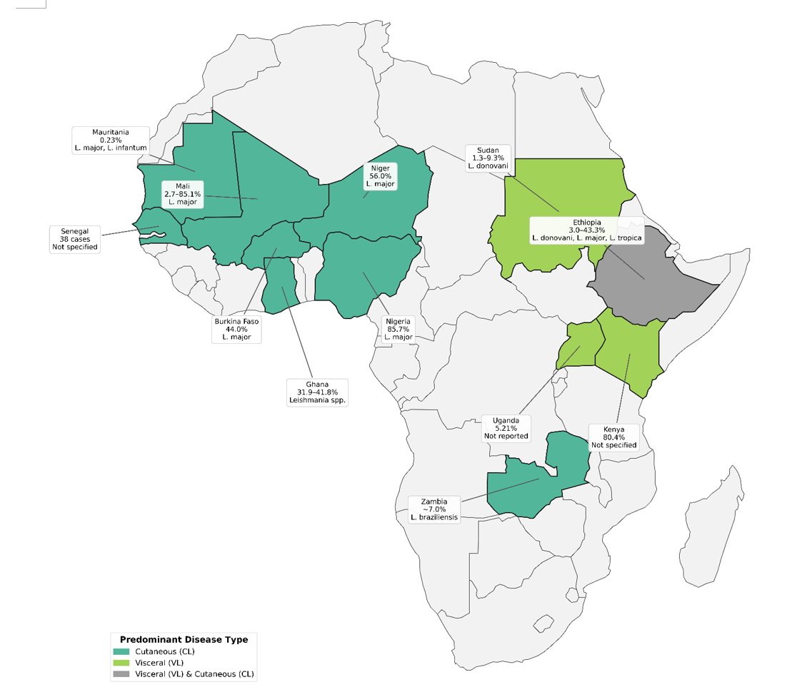
Leishmaniasis remains a major neglected tropical disease in Sub-Saharan Africa (SSA), manifesting predominantly as cutaneous leishmaniasis (CL) and visceral leishmaniasis (VL). Between 2010 and 2025, epidemiological investigations across Sub-Saharan Africa have revealed substantial heterogeneity in transmission intensity, affected populations, parasite species distribution, and comorbidities (Table 1 and Figure 1). Both community-based surveys and hospital-based studies demonstrate that leishmaniasis persists as a focal yet highly burdensome disease, driven by ecological, occupational, and socio-political determinants.
Cutaneous Leishmaniasis in West Africa
West Africa demonstrates sustained transmission of CL, primarily due to Leishmania major. In Burkina Faso, a retrospective hospital-based series (2012–2016) reported 44.0% parasitological positivity among suspected cases, with 96 confirmed infections in Ouagadougou [12]. This remarkably high proportion of confirmed infections among suspected cases carries profound implications for clinical practice. It suggests that healthcare providers in the region must maintain a high index of suspicion for cutaneous leishmaniasis when evaluating patients with chronic, non-healing skin ulcers, and it underscores the urgent need to integrate rapid, point-of-care diagnostic tools into primary care to ensure timely and appropriate treatment. Similarly, in Mali, retrospective parasitological analysis confirmed 41 CL cases from archived slides across multiple regions, identifying L. major as the predominant species [8]. Community-based surveys further illustrate variable endemicity within Mali, where leishmanin skin test positivity ranged from 2.7% in Kolondieba to 85.1% in Diema, with PCR confirmation of lesions in selected cases[9]. These findings highlight intense localized transmission foci.
In Ghana, cross-sectional surveys in the Oti Region identified 31.9% PCR-confirmed infection among persons with skin ulcers and 41.8% exposure by leishmanin skin testing [7], indicating substantial subclinical and active transmission. Mauritania reported a lower prevalence of 0.23% in a prospective multicenter study in Nouakchott; however, molecular typing revealed mixed circulation of L. major (61.9%) and Leishmania infantum (23.8%), underscoring species diversity [24].
Emerging data from Niger demonstrated 56.0% confirmation among suspected CL cases in Zinder, again implicating L. major [10]. In northwestern Nigeria, molecular characterization confirmed CL in 85.7% of suspected cases in Katsina State, with L. major identified as the causative agent [11]. Senegal documented 38 CL cases in a dermatology referral hospital between 2010 and 2014, reflecting continued though likely underreported transmission [25]. Collectively, CL epidemiology in West Africa is characterised by focal high-prevalence clusters, dominant transmission of L. major, and under-recognised community burden.
Visceral Leishmaniasis in East Africa and the Horn of Africa
East Africa, particularly Ethiopia, Sudan, Kenya, and Uganda, bears a disproportionate burden of VL, largely attributable to Leishmania donovani. Ethiopia remains the most extensively studied focus. Among children aged 4–15 years in Amhara Region, asymptomatic infection prevalence reached 9.9% [26]. A prospective cohort in northern Ethiopia documented 14.3% infection among volunteers in endemic areas [21]. Labour migrants in Kafta-Humera exhibited 12.5% seroprevalence, predominantly affecting males aged 15–34 years [3].
Hospital-based data reinforce high disease burden. At Metema Hospital, 21.6% of clinically suspected VL patients were confirmed positive [27]. In Oromia Region, retrospective cohort analysis (2013–2018) revealed 43.3% VL positivity among admitted patients, with substantial pediatric involvement and comorbid conditions including severe acute malnutrition, tuberculosis, malaria, and HIV [28]. Longitudinal data among HIV-infected individuals demonstrated asymptomatic VL prevalence of 12.8% in males and 4.2% in females, highlighting immunosuppression as a critical risk factor [29].
Migrant labourers constitute a particularly vulnerable population. Community-based studies in West Armachiho reported 9.6% prevalence [30]. A later prospective cohort demonstrated increasing prevalence over agricultural seasons, from 3.0% among first-time entrants at baseline to 36.4% among repeat laborers during follow-up [31], illustrating cumulative exposure risk in endemic lowland agricultural zones.
Conflict-related transmission has also emerged. In 2023, an outbreak of 1,050 CL cases among militia members in the Somali Region of eastern Ethiopia was attributed to Leishmania tropica, expanding the recognized epidemiological profile beyond traditional VL foci [32].
In Sudan, hospital-based surveillance in Gedarif identified significant coinfection rates among VL patients: 3.8% with HIV, 9.3% with malaria, and smaller proportions with hepatitis B and C [33]. These findings underscore the syndemic interaction between VL and other endemic infections.
Northern Kenya demonstrates a high VL burden. In Marsabit County, secondary data analysis revealed 80.4% positivity among confirmed VL cases evaluated between 2014 and 2017, with 83% of cases occurring in males [34]. In northeastern Uganda, community-based prevalence was 5.21%, with localized variation at the county level [6], indicating ongoing transmission outside historically recognized Ethiopian and Sudanese hotspots.
Leishmaniasis in Southern Africa
Leishmaniasis is relatively rare and under-documented compared to East Africa and the Horn of Africa, and the region is generally not considered a high risk for the acquisition of infection by either travelers or residents [35]. In recent years, there has been only one published report of a 15-year-old boy returning from South Africa with a cutaneous leishmaniasis ulcer and an identification of L. major [36]. The last reported cases of leishmaniasis in Namibia were over 3 decades ago [37]. There is a published report on a nurse who was diagnosed with cutaneous leishmaniasis three weeks after travelling to Botswana and visiting a game reserve. L. tropica was the implicated species in this case [13]. Although many species of Phlebotomine sandflies have been identified in Madagascar, there is no evidence to suggest that these species are capable of being vectors of Leishmania and transmitting the disease to humans. Because of this, the country is generally known to be leishmaniasis-free [14]. Although no case of human leishmaniasis had been reported in Zambia in over 4 decades, a study reporting cases of canine leishmaniasis caused by L. infantum in the country predicted the likely re-emergence of the disease [38,39]. In 2025, a pilot study using metagenomic next-generation sequencing (mNGS) on bio-banked nasopharyngeal swabs from COVID-19 patients in Lusaka, Zambia, detected molecular sequences of Leishmania (specifically matching L. braziliensis clade markers) with ~7% genomic detection rate [40]. Rather than confirmed clinical cutaneous leishmaniasis, this finding should be interpreted as molecular evidence of parasite exposure or low-level asymptomatic mucosal/respiratory colonization requiring further parasitological validation. This discovery calls for increased vigilance in monitoring leishmaniasis in the country. In addition, human cutaneous leishmaniasis has also been discovered in Angola after over 3 decades without a reported case [15].
This lack of information on the epidemiology of the disease in southern Africa is largely due to weak surveillance systems [14] and should not be interpreted as the absence of the disease. The historical perception that Southern Africa is free of leishmaniasis is likely a consequence of chronic under-detection rather than a true ecological absence of the parasite. Several intersecting factors contribute to this surveillance blind spot. First, there might be remarkably low clinical index of suspicion among healthcare providers in the region; because the disease is not traditionally considered endemic, clinicians rarely include it in their differential diagnoses. Consequently, cutaneous lesions are frequently misdiagnosed as other common tropical ulcers or bacterial skin infections, while the systemic symptoms of visceral leishmaniasis can easily be misattributed to malaria, disseminated tuberculosis, or HIV-related complications. Furthermore, the specialized diagnostic infrastructure required to confirm Leishmania infection is virtually absent in routine clinical settings across Southern Africa. Finally, because leishmaniasis is rarely integrated into national notifiable disease registries in these countries, the few cases that are accurately diagnosed often go unrecorded at the epidemiological level.
Methodological limitations and potential biases in current studies
While the data clearly show that leishmaniasis is a major burden in Sub-Saharan Africa, we have to be careful about how we interpret these numbers. The studies we currently rely on are incredibly diverse in their design, which naturally introduces a range of limitations and potential biases.
One major issue is that many regions rely heavily on hospital- or clinic-based sampling—for instance, the retrospective studies from Burkina Faso, Niger, and Senegal [10,12,25]. The problem here is selection bias: these studies only capture patients who are sick enough to need hospital care, and who have the money, transport, and access to get there. As a result, they likely overestimate the proportion of severe cases while completely missing the hidden reservoir of mild or asymptomatic infections out in the community. Conversely, community-based surveys (like those in Uganda, Ghana, and Mali) [6,7,9] give us a much better picture of local, active transmission. However, because they are often small and highly localized, we cannot easily scale their findings to estimate a true national prevalence. Additionally, studies that focus on specific high-risk groups, like migrant farm workers or military personnel in Ethiopia, give us great insight into occupational hazards, but they do not represent the everyday risk for the general public, including women and children.
Beyond who is being tested, how they are being tested makes comparing these studies exceptionally difficult. The definition of a ‘confirmed case’ varies depending on institutional diagnostic capacity, ranging from rapid diagnostic tests (RDTs) and skin tests to high-sensitivity PCR and direct microscopy. Consequently, variations in diagnostic sensitivity and specificity across tools introduce significant variance into reported prevalence rates, obscuring true epidemiological boundaries. Ultimately, this patchwork of different diagnostic tools and sampling methods means we have to exercise caution when conducting cross-country comparisons. The current literature most likely underreports the true, widespread burden of the disease across Sub-Saharan Africa, while at the same time artificially amplifying the specific hotspots where active research teams are operational.
Determinants of transmission and risk patterns
Across SSA, epidemiological patterns demonstrate consistent demographic and occupational risk gradients. Adult males engaged in farming, seasonal agricultural migration, or militia activities are disproportionately affected in VL-endemic zones [3,28,32]. Pediatric vulnerability remains evident in several Ethiopian cohorts [26,28]. HIV coinfection significantly amplifies infection risk and complicates disease dynamics [18,33].
CL transmission in West Africa appears strongly focal, often centred around rural settlements with ecological suitability for sandfly vectors. VL transmission in East Africa is associated with lowland agricultural expansion, population mobility, malnutrition, and immunosuppression. Beyond demographic and occupational risks, ecological and climatic variables serve as fundamental drivers of leishmaniasis transmission by strictly regulating both sandfly vector populations and reservoir host dynamics[41]. In Sub-Saharan Africa, seasonal rainfall and ambient temperature dictate vector abundance; warmer temperatures accelerate the development of Leishmania parasites within the sandfly gut, while specific soil moisture levels are required for breeding [42]. For instance, in East Africa, Phlebotomus vectors thrive in the unique microclimates of cracked ‘black cotton’ soils and termite mounds, which provide perfect, humid resting sites during extreme daytime heat [43].
Furthermore, anthropogenic ecological changes are rapidly shifting these traditional transmission boundaries [43]. Deforestation, unplanned urbanization, and the expansion of agricultural lands are altering local microclimates and pushing immunologically naïve human populations directly into sylvatic (wild) transmission cycles [44]. As climate change continues to drive erratic weather patterns, prolonged droughts, and shifting temperature gradients across the Sahel and the Horn of Africa, the geographic range of viable sandfly habitats is expected to expand, threatening to introduce the disease into previously unaffected or higher-altitude regions.
Coinfection and comorbidity of Leishmaniasis in Africa
Coinfection refers to the co-occurrence of leishmaniasis with other infections, such as HIV, TB, or malaria, often leading to compounded clinical manifestations, relapse rates, and death due to immunosuppression and drug pharmacokinetic changes. Malnutrition and other non-infectious comorbidities further complicate disease and treatment outcomes. The endemicity of Africa in nations such as Ethiopia, Sudan, South Sudan, and Kenya enhances these complications.
Leishmania-HIV coinfection is most common in East Africa, and Ethiopia has the highest occurrence rate, ranging from 20% to 30% [2]. Individuals with HIV are at increased risk of acquiring full-blown VL, having frequent relapses, and high mortality [2]. Antiretroviral therapy (ART) reduces these risks by slowing disease progression and improving survival [18]. In 2022, the World Health Organization published region-specific treatment guidelines for VL-HIV coinfection in East Africa. It recommended combination therapy with liposomal amphotericin B (LAmB) and miltefosine to enhance outcomes [2]. A cohort study from Ethiopia showed low exposure to antileishmanial drugs in HIV-VL patients, where the Cmax of amphotericin B was twice as low as in non-VL patients and exposure to miltefosine was 35% less in non-HIV VL cases, with a possibility of dose optimization needed for controlling elevated relapse rates [45]. ARV levels remained unaltered with antileishmanial therapy, though subtherapeutic efavirenz levels in a few patients underscore the importance of monitoring [45]. Earlier studies in 2010 indicated an increasing rate of VL-HIV coinfections in East Africa and, even more so, in Ethiopia amidst a reduction in the overall HIV cases, with clinical presentation showing atypical presentation and poorer treatment outcomes [16]. By 2016, comprehensive reviews evaluating VL-HIV co-infection across endemic areas highlighted atypical clinico-epidemiological presentations, elevated mortality risk, poor quality of life, and ongoing management challenges [46]. In highly epidemic areas like North-West Ethiopia, 19–41% of VL patients are HIV co-infected [17]. Diagnostic and treatment health care remains limited to few centers with limited clinical expertise [46].
VL-malaria coinfection presents a concern in overlapping endemic areas like West Pokot County, Kenya, where shared vectors and environmental determinants foster risks of comorbidity [20]. An observational study protocol would seek to contrast symptomatology, immunological interactions, and household determinants among mono- and coinfections and possibly inform targeted control measures [20].
In vitro studies of aminoquinoline-chalcone hybrids demonstrated dual activity against L. major and L. donovani (responsible for CL and VL, respectively), suggesting potential for treating comorbidities with malaria, though in vivo validation is needed [47]
Parasite genotype significantly influences VL mortality in Brazil, explaining up to 83% of mortality risk (heritability h² = 0.83 ± 0.17) based on L. infantum isolates from northeastern Brazil, with copy number variants (CNVs) in genes like LINF_070018000 associated with poor outcomes [48]. This genetic variation also correlates with comorbidities such as HIV and bacterial coinfections, emphasizing parasite factors in disease severity [48].
TB-VL coinfection, often in the context of HIV, represents an evidence gap, with prevalence estimates ranging from 5.7% to 29.7% in routine care settings, but diagnostic approaches are inconsistent [19]. Systematic TB screening in VL-HIV patients is advocated to assess clinical impact and enable early intervention, as TB contributes to high mortality in this group [19]. Malnutrition exacerbates progression to active disease, with diets deficient in protein, iron, vitamin A, and zinc heightening risks [2,18]. Post-kala-azar dermal leishmaniasis (PKDL), a sequel to VL comorbidity, is seen in 5-10% of the East African cases, serving as a reservoir for transmission and an earlier event in HIV-co-infected individuals [49].
Collectively, coinfections like HIV-VL and VL-malaria, comorbidities of Post-kala-azar dermal leishmaniasis (PKDL) and malnutrition, sustain a poor prognosis in Africa. Novel guidelines and pharmacokinetics demonstrate the need for tailored treatments, while diagnostic gaps in TB require heightened surveillance. Literature has documented shifting away from descriptive epidemiology to predictive modelling and region-specific interventions, reflecting novel challenges in co-endemic regions.
It is important to emphasize that the high rates and clinical severity of these coinfections do not happen in a vacuum; they are the direct result of a broader syndemic reality. The way leishmaniasis, HIV, malaria, and tuberculosis overlap and worsen one another is heavily driven by shared social, economic, and environmental factors. Issues like extreme poverty, conflict-driven displacement, severe malnutrition, and overlapping ecological vectors create the perfect storm, allowing these diseases to cluster and thrive in the most vulnerable populations.
Species distribution
Species distribution in SSA demonstrates geographic stratification. Leishmania major predominates in West African CL foci, including Burkina Faso, Mali, Niger, Nigeria, and Mauritania [8,10–12]. Leishmania donovani drives VL transmission in Ethiopia, Sudan, Kenya, and Uganda [6,26,27,34]. The emergence of Leishmania tropica in eastern Ethiopia during conflict highlights shifting epidemiological landscapes [32]. Mixed circulation, including L. infantum, has been documented in Mauritania [24]. Leishmania aethiopica is predominantly localized to the highlands of Ethiopia and Kenya, serving as a key agent of localized and diffuse cutaneous leishmaniasis [50,51].
Molecular and genetic aspects of Leishmaniasis: Strains, implications, and drug resistance gene studies
Molecular and genetic studies have vastly expanded our understanding of Leishmania strains by detailing virulence mechanisms and drug resistance. However, the true value of these molecular insights lies in their translational relevance to epidemiology and public health. For highly endemic regions in Africa, identifying the genetic markers of L. donovani (VL), L. major (CL), and L. infantum (VL/CL) is no longer just an academic exercise; it is the foundation of modern genomic surveillance. By tracking specific genetic adaptations, public health programs can map transmission networks, monitor the emergence of drug-resistant strains in real-time, and proactively adapt treatment guidelines before widespread clinical failures occur. Genome-wide screening identified 22 phosphatidylinositol-specific phospholipase C (PI-PLC) genes in L. infantum and L. major, conserved for parasite survival and immune evasion [52]. Differential expression in antimony-resistant L. infantum strains suggests PI-PLC’s role in drug susceptibility, positioning these genes as potential molecular markers for resistance monitoring [52]. Phylogenetic analysis classified PI-PLC into subfamilies, highlighting evolutionary conservation and implications for cross-strain therapies [52].
In L. donovani, dithiol glutaredoxin 1 (LdGrx1) maintains redox homeostasis and is upregulated in amphotericin B (Amp-B)-resistant strains, correlating with higher IC50 values, elevated thiols, and reduced reactive oxygen species (ROS) [53]. This cytosolic protein’s role in oxidative stress management implies it as a virulence factor enabling survival under drug pressure [53]. Whole genome sequencing (WGS) of multiple L. donovani clinical isolates from Africa and beyond has provided insights into population structure and drug resistance mechanisms, revealing genetic heterogeneity and within-host evolution [54–56]. These studies show that resistance in Brazilian Leishmania infantum isolates is linked to inherent traits observed in pretreatment isolates, specifically a deletion in the miltefosine sensitivity locus (MSL). This suggests a natural resistance mechanism present before miltefosine use in Brazil, with a significant correlation between MSL absence and higher IC50 values (P = 0.0301), predicting treatment failure with 92% sensitivity and 78% specificity [57]. Post-genomic approaches from 2013 onward have tackled resistance by expanding beyond single genes to whole-genome analyses, identifying novel markers [58]. From an epidemiological standpoint, these genomic insights are critical for field monitoring. For instance, discovering that resistance is linked to inherent traits like the miltefosine sensitivity locus (MSL) deletion means that this locus can be used as a predictive molecular marker in clinical settings [57]. Rather than waiting for a patient to fail therapy, screening for such genetic variants could allow clinicians to predict treatment failure and switch therapies early, thereby reducing mortality and preventing the further transmission of resistant strains in the community.
Refractory ML, often due to L. braziliensis (relevant for comparative insights), involves overexpression of heat shock protein 70 (HSP70) and ATPase, impairing host hydrogen peroxide production and promoting parasite persistence despite low burdens[59]. This results in treatment failures in various drugs, highlighting drug-resistant strain genetic adaptations [59]. Molecular mechanisms of drug resistance in Leishmania spp. include genomic, transcriptional, and translational control, with a review in 2024 highlighting specific mutations linked with antileishmanial drug resistance [60]
Subtractive genomics identified glutamate dehydrogenase (GDH) as a conserved Leishmania drug target with the closest phylogenetic evolutionary relationship revealed [61]. Molecular docking and simulations showed GW5074’s strong binding to GDH, suggesting it as a candidate inhibitor to combat AMR [61]. Genomic plasticity drives resistance, with reviews emphasizing the paradigm of intracellular parasite survival through gene expression changes in antimony-resistant strains [62].
Genetic engineering advances, including CRISPR/Cas and Cre-loxP systems, enable the creation of live attenuated vaccines by disrupting pathogenesis genes in L. major, offering long-term protection against virulent strains [63]. These tools overcome classical attenuation limitations, with implications for strain-specific immunity. Next-generation sequencing for drug resistance analysis in L. donovani has revealed natural population heterogeneity under treatment pressure, with implications for field monitoring [64].
Antimony-resistant L. major MON-25 maintains its phenotype post-passage through Phlebotomus papatasi, with resistance genes (e.g., LmTDR1, LmMRPA) unaltered, indicating potential field transmission despite reduced late-stage infectivity [65]. This highlights vector-parasite interactions in perpetuating resistant strains.
Ultimately, the genetic plasticity of Leishmania spp. drives not only drug resistance but also the parasite’s ability to adapt to different vectors and ecological niches across Sub-Saharan Africa. The genes driving resistance and virulence, such as PI-PLC, LdGrx1, HSP70, and GDH, should be integrated into regional epidemiological monitoring as standard biomarkers. Transitioning these molecular findings from the laboratory to the field will be essential for developing rapid point-of-care diagnostics, mapping the spread of resistant phenotypes, and designing targeted control strategies capable of outpacing the parasite’s evolution.
Diagnosis of Leishmaniasis
In Africa, laboratory confirmation of leishmaniasis has traditionally depended on a combination of clinical suspicion and parasitological verification. In endemic settings, prolonged fever, weight loss, and hepatosplenomegaly commonly raise suspicion of visceral disease, whereas chronic or ulcerative skin lesions suggest cutaneous involvement. Confirmation has historically relied on light microscopy of stained tissue aspirates or smears using Romanowsky-based stains such as Giemsa or Wright–Giemsa to visualize intracellular amastigotes [18,66]. Additional parasitological approaches include in vitro cultivation of promastigotes and experimental inoculation into animal models, typically mice, to observe disease progression. Although these methods remain accessible and relatively inexpensive, they generally permit identification only at the genus level and do not reliably differentiate species [66,67].
Diagnostic capacity in Sub-Saharan Africa has expanded with the adoption of serological assays, rapid diagnostic platforms, and molecular tools. Immunological methods detect parasite antigens or host antibodies and include latex agglutination, the Montenegro skin test, direct agglutination testing, indirect immunoblotting, hemagglutination assays, immunofluorescence antibody testing, ELISA, and immunochromatographic assays. The latex agglutination test uses antigen-coated particles and demonstrates high sensitivity, though specificity varies [68]. The Montenegro test, based on delayed-type hypersensitivity, is inexpensive and sensitive but may be negative in early visceral infection [69]. Direct agglutination testing employs trypsin-treated promastigotes and offers favorable sensitivity and specificity; however, seropositivity can persist after clinical cure [70,71]. Immunoblotting enhances diagnostic precision by detecting serum antibodies against separated parasite antigens [72]. Indirect hemagglutination assays use sensitized erythrocytes and show variable performance depending on host immune status [73]. Immunofluorescence assays are accurate but require specialized infrastructure [66]. ELISA remains widely applied because of its high diagnostic performance, although outcomes depend on antigen selection [74–76]. The rK39-based immunochromatographic test has become a principal screening tool for visceral leishmaniasis due to its speed, simplicity, and suitability for field deployment. Nonetheless, sensitivity differs geographically; reduced performance in East Africa has been linked to genetic variability among circulating strains of Leishmania donovani, limiting uniform applicability [70,71,76].
Molecular diagnostics have further strengthened detection and characterization. Polymerase chain reaction remains the most widely implemented platform, enabling amplification of parasite nucleic acids from blood, bone marrow, or tissue biopsies with superior sensitivity and species-level discrimination. It is particularly useful in relapse confirmation and epidemiological investigations. However, cost, technical requirements, and limited infrastructure restrict routine implementation in many Sub-Saharan African settings [73]. Enhanced variants have been introduced to overcome limitations of conventional PCR. Nested and semi-nested PCR improve analytical sensitivity but increase contamination risk [22,77]. Real-time PCR allows simultaneous amplification and quantification, though species discrimination may be limited in co-endemic regions [77,79]. Multiplex PCR targets multiple loci concurrently, increasing efficiency but also cost and complexity [80]. NASBA amplifies RNA targets and provides high sensitivity, yet does not reliably differentiate species [23]. For detailed genotyping and epidemiological surveillance, PCR-RFLP and AFLP enable strain-level discrimination through restriction analysis of amplified fragments, supporting detection of low parasite burdens and transmission mapping [81,82].
Loop-mediated isothermal amplification represents a promising field-adapted molecular option because it operates without thermal cycling and requires minimal equipment [83–85]. Despite technological advances, diagnostic challenges persist. The broad clinical spectrum of cutaneous disease, species diversity, and the co-endemic presence of other febrile illnesses such as tuberculosis, typhoid, and malaria contribute to delayed or missed diagnoses in many endemic regions.
While molecular diagnostics offer unparalleled sensitivity, their real-world application in Sub-Saharan Africa faces significant logistical hurdles. The routine use of PCR and targeted sequencing requires a strict cold chain for reagents, stable electricity for thermocycling, and highly trained personnel for complex DNA extractions. Because these resources are rarely available in the rural primary care clinics where the disease burden is highest, these powerful tools remain largely confined to central reference laboratories. This stark divide between diagnostic capacity and clinical need highlights the urgency for field-ready alternatives, such as freeze-dried LAMP assays, that can withstand resource-limited settings.
Advances in treatment of Leishmaniasis
Therapeutic approaches to leishmaniasis across Africa reflect regional species distribution and resource availability. In Ethiopia, localized cutaneous disease caused by Leishmania aethiopica is managed with topical paromomycin, thermotherapy, cryotherapy, and intralesional or systemic meglumine antimoniate [50]. Although intravenous pentamidine and amphotericin B formulations, including liposomal preparations, demonstrate efficacy, their use is limited [86,87]. In Kenya, intralesional antimonials are the predominant treatment modality [51]. High-dose intravenous sodium stibogluconate is effective for single lesions due to L. aethiopica, though relapses have been reported with L. tropica infections [88]. Heat therapy has also been explored but with documented recurrence. In Sudan, intravenous meglumine antimoniate and oral ketoconazole have shown clinical benefit for cutaneous and mucosal disease [88].
Across West Africa, particularly in Senegal, Mauritania, and Niger, intralesional meglumine antimoniate is commonly used against L. major [25,88]. Oral ketoconazole and metronidazole are also employed in some contexts [88]. In Ghana, cases linked to recently characterized species are frequently managed with traditional plant-based remedies or improvised thermotherapy [7]. In Cameroon, where L. major predominates, topical amphotericin B deoxycholate and oral metronidazole have been reported as effective options [88]. Data from Southern Africa remain limited, though topical paromomycin, oral fluconazole, and occasionally surgical intervention have been described, despite surgery not being generally recommended for cutaneous disease [36,88].
Rising treatment failure and emerging drug resistance have stimulated exploration of alternative strategies. Combination regimens aim to shorten treatment duration, lower toxicity, reduce relapse, and delay resistance development [89]. Field-adapted combination regimens combining short-course first- and second-line therapeutics have demonstrated substantial clinical efficacy. In Sudan, clinical evaluations of combined liposomal amphotericin B, miltefosine, and paromomycin demonstrated high cure rates and improved safety profiles in patients with post-kala-azar dermal leishmaniasis (PKDL) [90]. In immunocompromised individuals co-infected with HIV, combined miltefosine and liposomal amphotericin B has shown encouraging outcomes [90].
Drug repurposing offers an efficient strategy by evaluating approved agents for antileishmanial activity. Amphotericin B, miltefosine, azole antifungals, and sertraline have exhibited leishmanicidal effects [91,92].
Nanotechnology-based delivery systems have introduced targeted therapeutic approaches, particularly given the intracellular localization of parasites within macrophages. Liposomal and polymeric nanoparticle platforms enhance drug solubility, cellular uptake, and targeted delivery while reducing systemic toxicity [93,94]. Nanoparticle-mediated delivery has been associated with parasite killing mechanisms such as oxidative stress induction, mitochondrial membrane disruption, and inhibition of trypanothione reductase [95].
Natural products remain a significant source of candidate compounds. Extracts from Asparagus gracilis, Stellaria media, Sida cordata, and Jurinea dolomiaea have shown activity against Leishmania tropica [96–98]. Brazilian plant species, including Dipteryx alata, Hymenaea stignocarpa, and Jacaranda cuspidifolia have yielded active fractions against L. amazonensis [99,100]. Essential oils derived from medicinal plants, such as Artemisia annua, demonstrate notable leishmanicidal activity against visceral disease [101]. Alkaloids such as solamargine and solasonine from Solanum lycocarpum have surpassed standard drugs in certain models [102]. Flavonoids, lignans, coumarins, quinones, and terpenoids have also shown efficacy, including quercetin and apigenin from Kalanchoe pinnata, dehydrodieuginol, coumarins from Calophyllum brasiliense, and terpenoids such as artemisinin, (-)-α-bisabolol, ursolic acid, and oleanolic acid, with documented in vivo lesion reduction [101,103–106]. Marine-derived terpenoids from Dendrilla membranosa and Plumarella delicatissima and nanoformulated plant compounds further expand the therapeutic pipeline [107,108].
While historical and experimental therapies remain part of the literature, clinical practice in Sub-Saharan Africa is guided by specific regional protocols. Table 2 outlines the current World Health Organization (WHO) recommended therapeutic regimens, distinguishing between first-line standard of care and second-line alternatives based on the clinical presentation [2].
Challenges with control and prevention of leishmaniasis in Africa
Control and prevention of leishmaniasis in Africa remain difficult because transmission is sustained by a combination of patient, health-system, and environmental factors. Limited public awareness in endemic communities reduces early care-seeking and weakens the uptake of preventive measures, while effective control also depends on sustained community engagement and local understanding of reservoir and vector ecology [109–111].
Socioeconomic deprivation is a major barrier to prevention. Poor housing, inadequate sanitation, and unsafe waste disposal create favourable conditions for sandfly breeding and human exposure, and these risks are intensified by overcrowding and poverty. Nutritional deficiencies, including protein-energy undernutrition and shortages of iron, zinc, and vitamin A, further increase susceptibility and may worsen disease severity [1,110,112–116].
Human behaviour also contributes to transmission. Sleeping outdoors or on the ground, especially in endemic settings, increases contact with sandflies and raises infection risk. In addition, immunosuppressive conditions, particularly HIV infection, complicate diagnosis and management by increasing the risk of severe disease, relapse, and death, and by reducing the reliability of some diagnostic tests[116–118].
Health-system constraints further undermine control efforts. Leishmaniasis often mimics other febrile, dermatological, or systemic illnesses, leading to misdiagnosis and delayed treatment, especially where confirmatory laboratory capacity is limited [110,119]. Access to effective drugs is also restricted by cost, toxicity, and the need for parenteral administration, while emerging drug resistance increases the risk of treatment failure [92,110,120,121].
Environmental and structural changes add another layer of complexity. Climate change, population displacement, unplanned urbanization, conflict, and land-use change alter sandfly habitats and increase human-vector contact, creating new transmission foci [122,123]. Vector and reservoir control remains difficult because of parasite diversity, ecological heterogeneity, and the limited feasibility and sustainability of existing control measures.
Because health system capacities vary drastically across Sub-Saharan Africa, overcoming leishmaniasis requires moving away from a “one-size-fits-all” approach and adopting a context-specific, tiered strategy:
Ultimately, these tiered efforts must be supported by strong cross-border collaboration to effectively manage transmission among migrant and displaced populations.
Overall, effective prevention will require integrated strategies that combine community education, strengthened surveillance, improved diagnosis, affordable treatment access, and context-specific vector control.
Leishmaniasis in Sub-Saharan Africa is far more widespread, heterogeneous, and operationally complex than previously appreciated. Evidence from West, East, and Southern Africa shows that transmission is shaped by species diversity, ecological change, human mobility, conflict, poverty, and overlapping infections, with CL and VL posing distinct but interconnected public health challenges.
Taken together, the findings from this review underscore the need to move beyond passive recognition of the disease toward integrated, region-specific control strategies. Strengthening surveillance, expanding access to accurate species-level diagnostics, improving case management for HIV and other coinfections, and ensuring availability of safer and more effective treatments are essential to reducing missed diagnoses, relapse, and mortality.
Future progress will depend on linking molecular epidemiology with field-based public health action. Investments in vector ecology, reservoir studies, resistance monitoring, and implementation research will be critical for developing sustainable interventions and for closing the large knowledge gaps that continue to obscure the true burden of leishmaniasis across the region.
What is already known about the topic
What this study adds
Conceptualization: Oyetunde Timothy Oyeyemi
Writing – Original Draft: Oyetunde Timothy Oyeyemi, Adedayo Oluwadamilola Adesida, Blessing Mercy Babalola1, Praise Irabor, Tobi Emmaneul Eludipo
Writing – Review & Editing: Oyetunde Timothy Oyeyemi, Adedayo Oluwadamilola Adesida, Blessing Mercy Babalola1, Praise Irabor, Tobi Emmaneul Eludipo
Abbreviations
ACT: Artemisinin-based Combination Therapy
AFLP: Amplified Fragment Length Polymorphism
Amp-B: Amphotericin B
ART: Antiretroviral Therapy
ARV: Antiretroviral
CL: Cutaneous Leishmaniasis
CNV: Copy Number Variant
CRISPR: Clustered Regularly Interspaced Short Palindromic Repeats
DAT: Direct Agglutination Test
DNA: Deoxyribonucleic Acid
ELISA: Enzyme-Linked Immunosorbent Assay
GDH: Glutamate Dehydrogenase
HIV: Human Immunodeficiency Virus
HSP70: Heat Shock Protein 70
IC50: Half Maximal Inhibitory Concentration
KAtex: Latex Agglutination Test for Kala-azar
LAMP: Loop-Mediated Isothermal Amplification
LAmB: Liposomal Amphotericin B
MSL: Miltefosine Sensitivity Locus
NASBA: Nucleic Acid Sequence-Based Amplification
PCR: Polymerase Chain Reaction
PCR-RFLP: Polymerase Chain Reaction Restriction Fragment Length Polymorphism
PI-PLC: Phosphatidylinositol-Specific Phospholipase C
PKDL: Post-Kala-Azar Dermal Leishmaniasis
ROS: Reactive Oxygen Species
rK39: Recombinant Kinesin 39 Antigen
RNA: Ribonucleic Acid
SSA: Sub-Saharan Africa
TB: Tuberculosis
VL: Visceral Leishmaniasis
VL-HIV: Visceral Leishmaniasis and Human Immunodeficiency Virus Coinfection
WHO: World Health Organization
WGS: Whole Genome Sequencing
Table 1: Epidemiology of Leishmaniasis in Sub-Saharan African Countries (2010–2025)
| S/No | Country | Prevalence | Type of Study | Year of Data Collection | Population Type | Location | Age | Type of Leishmaniasis | Species of Leishmania | Reference |
|---|---|---|---|---|---|---|---|---|---|---|
| 1 | Burkina Faso | 44.0% parasitological positivity among suspected cases; 96 confirmed cases | Retrospective hospital case series | 2012–2016 | Patients with suspected CL | Ouagadougou | ~30.8 years average among those with age reported | CL | Leishmania major | [12] |
| 2 | Ghana | 31.9% among persons with skin ulcers detected by PCR; 41.8% exposure by leishmanin skin test | Cross-sectional community surveys | 2018 | Community residents and persons with skin ulcers | Oti Region (Ashiabre, Keri, Sibi Hilltop) | Not reported | CL | Leishmania spp. (PCR positive) | [7] |
| 3 | Mali | LST positivity 85.1% (Diema), 24.6% (Kolokani), 2.7% (Kolondieba) with PCR-confirmed lesions in some cases | Population survey using leishmanin skin test and PCR confirmation | 2012–2016 (community survey) | General community residents in multiple districts | Districts: Diema, Kolokani, Kolondieba | Median age: 23 years | CL | Leishmania major | [9] |
| 4 | Mauritania | 0.23% | Prospective multicenter descriptive epidemiological and clinico-biological study | 2016–2017 | Patients presenting with suspected cutaneous leishmaniasis lesions at two public hospitals in Nouakchott | Nouakchott | 23 years | CL | Leishmania major in 61.9% of cases; Leishmania infantum detected in 23.8% of cases | [24] |
| 5 | Niger | 56.0% (33 confirmed CL among 59 suspected) | Clinic case series | 2023 | Patients with suspected CL lesions | Zinder area | Not reported | CL | Leishmania major | [10] |
| 6 | Nigeria | 85.7% (6/7 suspected with molecular confirmation of CL lesions) | Molecular case series | 2024 | Patients with suspected CL lesions | Dutsin-Ma LGA (Katsina State) | Not reported | CL | Leishmania major | [11] |
| 7 | Senegal | 38 cases reported | Retrospective hospital case series | 2010–2014 | Patients with CL presenting to dermatology department | Aristide Le Dantec University Hospital Dermatology, Dakar | ~25 years | CL | Not specified | [25] |
| 8 | Ethiopia | DAT – 12.5%; Seroprevalence – 32.5% | Cross-sectional study | October 2013 | Labour migrant workers | Kafta-Humera lowlands, Northwest Ethiopia | Range 14–61 years (79.9% aged 15–34) | Visceral | Leishmania donovani | [3] |
| 9 | Ethiopia | 21.6% | Cross-sectional study | January 2014–June 2014 | Clinically suspected VL patients, predominantly males (87%), including age groups under 5 years and 15–29 years, with farmers (69.5%) and rural residents (60.9%) | Metema Hospital, North Gondar Administrative Zone, Amhara region, Northwest Ethiopia | 28.1 years (SD 11.8, range 2–78 years) | Visceral | Leishmania donovani | [27] |
| 10 | Ethiopia | 9.6% | Community-based cross-sectional study | October–December 2016 | Migrant laborers aged 15–65, predominantly 15–29 years (74.2%), from lowland areas (61.2%), engaged in sesame and sorghum harvesting | Agricultural camps, West Armachiho district, Northwest Ethiopia | 15–65 years | Visceral | Leishmania donovani | [30] |
| 11 | Ethiopia | 12.8% in males and 4.2% in females | Prospective cohort | 2015–2016 | HIV-infected individuals | Metema District Hospital, North-West Ethiopia | Median 38 years (IQR 30–45) | Visceral | Leishmania donovani | [29] |
| 12 | Ethiopia | 43.3% | Retrospective cohort study | September 2013–September 2018 | VL patients, including children (51.6%), with concurrent conditions (e.g., severe acute malnutrition 27.1%, tuberculosis 6.4%, malaria 6.4%, HIV 1.1%) | Negelle General Hospital, Yabello General Hospital, Ginnir General Hospital, Oromia region, Ethiopia | Not reported | Visceral | Leishmania donovani | [28] |
| 13 | Ethiopia | 3.0% (first comers at T1), 22.1% (repeat comers at T1), 34.8% (first comers at T2), 36.4% (repeat comers at T2) | Prospective cohort | July–August 2019 (T1) to November 2019–January 2020 (T2) | Migrant workers | West Armachiho district, Northwest Ethiopia | Median 20 years | Visceral (asymptomatic) | Leishmania donovani | [31] |
| 14 | Uganda | 5.21% (95% CI: 3.15%–8.48%) in Moroto district, with county-level prevalence of 6.90% in Matheniko, 4.49% in Tepeth, and 3.61% in Moroto Municipality | Cross-sectional study | Not reported (published 2025) | General community population | Moroto district, Northeastern Uganda | Not reported | Visceral | Not reported | [6] |
| 15 | Ethiopia | Not reported (outbreak with 1050 cases) | Cross-sectional study | 2023 | Male militia members | Somali Region, Eastern Ethiopia | Not reported | Cutaneous | Leishmania tropica | [32] |
| 16 | Sudan | VL/HIV co-infection: 3.8%; VL/HBV co-infection: 1.9%; VL/HCV co-infection: 1.3%; VL/Malaria co-infection: 9.3%; VL/HIV/HBV/HCV/Malaria co-infection: 1.9% | Retrospective, hospital-based study | January 2013–June 2014 | Confirmed VL patients, predominantly males (75.7%), farmers (56.2%), rural residents (74.4%) | Gedarif Teaching Hospital, Eastern Sudan | 31.4 years | Visceral | Leishmania donovani | [33] |
| 17 | Kenya | 80.4% | Secondary data analysis and frequency-matched case-control study | 1st January 2014–11th July 2017 | Confirmed VL cases (83% males) and controls (age-matched, VL-negative), including children under 5 years | Marsabit County Referral Hospital and Laisamis Mission Hospital, Northern Kenya | Not reported | Visceral | Not specified | [34] |
| 18 | Zambia | ~7.0% | A metagenomic next-generation sequencing-based pilot study was conducted on bio-banked nasopharyngeal samples | November 2020–February 2021 | COVID-19 referral centers in Lusaka, Zambia, the University Teaching Hospital (UTH) and Levy Mwanawasa University Teaching Hospital (LMUTH) | Lusaka | ≥18 years | Molecular detection / Exposure (Nasopharyngeal mNGS) | L. braziliensis | [40] |
Note: This table summarizes population-level epidemiological surveys, hospital cohorts, and community-based molecular studies. Singular clinical case reports and isolated travel-associated infections have been excluded from this summary to maintain comparability across prevalence data. CL – cutaneous leishmaniasis; VL – visceral leishmaniasis; DAT – direct agglutination test; HBV – hepatitis B virus; HCV – hepatitis C virus.
Table 2: Recommended treatment regimens for leishmaniasis in Sub-Saharan Africa
| Clinical Form | Primary Regional Focus | First-Line Recommended Treatment | Second-Line / Alternative Treatment |
|---|---|---|---|
| Visceral Leishmaniasis (VL) | East Africa | Sodium Stibogluconate (SSG) + Paromomycin (PM) combination | Liposomal Amphotericin B (L-AmB) or Miltefosine |
| Cutaneous Leishmaniasis (CL) – Uncomplicated | West & Southern Africa | Local therapy (Intralesional antimonials, cryotherapy, or thermotherapy) | Systemic antimonials or oral Fluconazole |
| Cutaneous Leishmaniasis (CL) – Complex/Multiple lesions | Cross-regional | Systemic antimonials (SSG or Meglumine antimoniate) | Miltefosine or Liposomal Amphotericin B (L-AmB) |